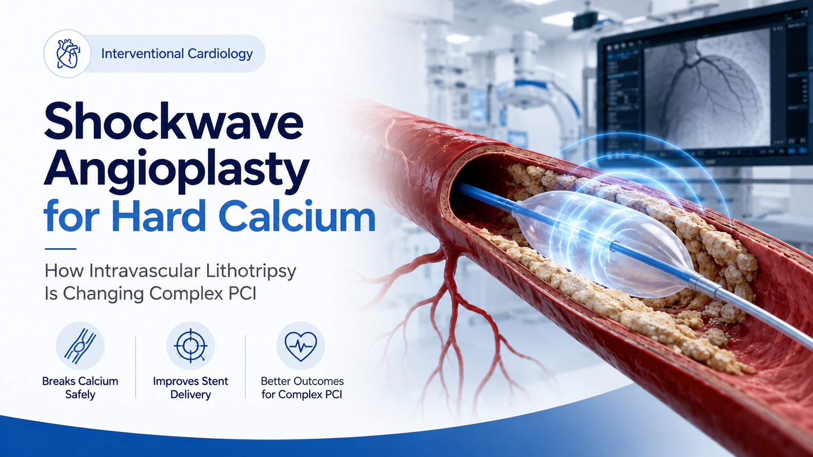
Most people understand angioplasty as a procedure in which a balloon is used to open a blocked heart artery and a stent is placed to keep the artery open.
But not every heart blockage behaves the same way.
Some coronary blockages contain a large amount of calcium, making the artery extremely rigid. Instead of behaving like a flexible blood vessel, the artery may become almost like a hard pipe.
In these situations, simply inflating a conventional angioplasty balloon may not adequately expand the blockage.
The balloon may fail to open completely. More importantly, if the calcium underneath the blockage is not properly modified, the stent may also fail to expand fully.
This is where advanced calcium-modification technologies such as Intravascular Lithotripsy (IVL) have changed the way cardiologists approach complex coronary angioplasty.
IVL is commonly referred to as Shockwave Angioplasty because it uses controlled acoustic pressure waves inside the artery to fracture calcium before a stent is implanted.
The concept is similar to the shockwave technology used to break kidney stones, although coronary IVL is specifically designed for use inside blood vessels.
Current interventional cardiology experience shows that IVL can fracture both superficial and deeper calcium, making the artery more compliant and allowing better balloon and stent expansion.
Coronary artery disease develops when cholesterol-containing plaque gradually builds up within the arteries supplying blood to the heart.
Over time, some plaques can become heavily calcified.
Calcium within the coronary arteries is particularly common in patients who are:
Calcification itself does not mean that angioplasty cannot be performed.
The challenge is how much calcium is present, where it is located, how thick it is and how extensively it surrounds the artery.
Severe circumferential or thick calcium can prevent a coronary stent from opening to its intended diameter.
During PCI — Percutaneous Coronary Intervention — a stent needs to expand properly and sit firmly against the wall of the coronary artery.
Think of a normal artery as relatively flexible.
When an angioplasty balloon is inflated, the vessel can expand sufficiently to accommodate the stent.
A severely calcified artery is different.
This can create several technical problems during PCI.
Severely calcified and narrow lesions may make it difficult to advance balloons, stents or imaging catheters through the blockage.
Even a high-pressure balloon may develop a visible narrowing in its centre because the calcium refuses to yield.
This is one of the most important concerns.
If the artery does not expand adequately, the stent implanted inside it may also remain partially underexpanded.
Severe coronary calcification is associated with greater procedural complexity and can contribute to problems such as stent underexpansion and malapposition.
Therefore, in heavily calcified coronary disease, treating the calcium is often an important step before the final stent is implanted.
Intravascular Lithotripsy, or IVL, is a catheter-based technology designed specifically to modify calcium inside blood vessels.
During the procedure, a specially designed balloon catheter containing lithotripsy emitters is positioned across the calcified blockage.
The balloon is gently inflated.
The system then generates short pulses of acoustic pressure energy.
These pressure waves travel through the soft tissue of the artery and interact with the rigid calcium.
Instead of simply forcing the artery open with very high balloon pressure, IVL attempts to create microfractures within the calcium.
The calcium does not disappear.
Instead, it becomes fractured and more flexible.
Once the calcium has been modified, the artery can expand more effectively when a balloon is inflated and a stent is subsequently implanted.
IVL typically operates with relatively low balloon pressures while acoustic energy modifies both superficial and deeper calcium.
The treatment can be understood in a few steps.
The cardiologist first identifies the location and severity of the coronary blockage using coronary angiography.
If significant calcification is present, additional imaging may be considered.
In complex cases, technologies such as:
IVUS – Intravascular Ultrasound
or
OCT – Optical Coherence Tomography
can provide a detailed view of the artery from inside.
They can help determine:
Intravascular imaging has become increasingly important in planning and optimizing complex PCI, particularly when coronary calcification is substantial.
A specialised IVL balloon catheter is advanced over a coronary guidewire until it reaches the calcified blockage.
The balloon is positioned precisely across the calcium.
The balloon is gently inflated.
The system then delivers a series of controlled acoustic pressure waves.
These waves pass through soft tissue but interact with rigid calcium, producing fractures within the calcified plaque.
The process can be repeated along different segments of the lesion when necessary.
Once the calcium has fractured sufficiently, the cardiologist reassesses the lesion.
A conventional or non-compliant balloon may then expand much more effectively.
After the artery has been properly prepared, the coronary stent is placed.
The aim is to obtain:
Imaging with IVUS or OCT may again be used to confirm the final result.
Regular balloon angioplasty works mainly by applying mechanical pressure against the blockage.
In mildly calcified disease, this may be enough.
With extremely hard calcium, however, increasing balloon pressure does not necessarily solve the problem.
IVL works differently.
Instead of depending only on balloon pressure, it uses acoustic energy to fracture calcium from within the vessel wall.
This can make a previously rigid segment of coronary artery more compliant.
No.
Both are important technologies used to manage severely calcified coronary blockages, but they work differently.
Rotational atherectomy — commonly called Rotablation — uses a rapidly rotating specialised burr to modify hard superficial calcium.
It can be especially useful when the blockage is extremely tight and balloons cannot cross or adequately engage the lesion.
IVL does not drill or mechanically remove calcium.
Instead, it sends acoustic pressure waves through the arterial wall to create calcium fractures.
It can affect both superficial and deeper calcium and does not rely on preferential contact between an atherectomy burr and one side of the artery.
There is no single device that is best for every calcified artery.
The choice depends on factors such as:
Sometimes rotational atherectomy may be required initially to make a very tight lesion crossable, followed by IVL to modify deeper calcium.
This combined approach is sometimes informally described as “RotaTripsy.”
Complex PCI therefore involves selecting the right tool for the specific anatomy rather than applying the same technique to every patient.
IVL may be considered when coronary angiography or intravascular imaging demonstrates significant calcification that is likely to interfere with optimal stent implantation.
Possible situations include:
Particularly when the calcium extensively surrounds the artery or is very thick.
If a properly selected balloon cannot satisfactorily expand a lesion, additional calcium modification may be required.
Calcification close to an important side branch can make PCI technically demanding.
One practical advantage of IVL in selected bifurcation procedures is that a side-branch guidewire can often remain in place during calcium modification.
Some carefully selected calcified left-main lesions may be treated using IVL-assisted PCI, although these are particularly complex procedures requiring detailed patient and anatomical evaluation.
Calcification is frequently one of the major challenges encountered during complex CTO PCI.
IVL can sometimes be incorporated into the calcium-modification strategy after the lesion has been successfully crossed.
Evidence for some of these more challenging uses continues to develop.
The purpose of angioplasty is not simply to get a stent across a blockage.
The stent must be properly implanted.
Imagine attempting to expand a metal framework inside a rigid concrete tube.
If the tube does not give way, the framework cannot completely expand.
The same principle applies to a severely calcified artery.
Calcium modification creates greater vessel compliance so that the stent can achieve better expansion.
This is important because stent underexpansion is an undesirable result following PCI.
The DISRUPT CAD programme has been central to the evaluation of coronary intravascular lithotripsy.
In the large DISRUPT CAD III study involving 431 patients with severely calcified coronary lesions, procedural success was reported in approximately 92% of patients.
The technology has subsequently been used across a broader range of calcified coronary anatomies in contemporary clinical practice.
However, IVL should not be viewed as replacing every other calcium-modification strategy.
Recent research presented in 2025 also suggests that appropriately selected cutting balloons and very-high-pressure balloon strategies can achieve comparable outcomes for some calcified lesions.
Current thinking is therefore increasingly focused on individualized, imaging-guided calcium modification rather than automatically choosing one device for every patient.
For appropriately selected patients, IVL offers several technical advantages.
The acoustic pressure waves can penetrate the vessel wall and fracture deeper calcium.
The procedure resembles techniques already familiar to interventional cardiologists.
Instead of relying entirely on very high pressure, the energy itself modifies the calcium.
Acoustic energy is transmitted around the vessel, which may be useful when calcium surrounds much of the artery.
By making the artery more compliant before stenting, IVL can facilitate more complete stent deployment.
IVL is increasingly incorporated into strategies involving:
However, the evidence base is stronger for some situations than others, and treatment must remain individualized.
Yes.
IVL is an important technology, but it is not suitable for every calcified lesion.
For example, if a blockage is so narrow that the IVL balloon cannot cross it, another technique may be necessary first.
Severely tortuous arteries can also make catheter delivery difficult.
Long areas of diffuse calcium may require treatment at several sites.
There may also be situations where cutting balloons, scoring balloons, high-pressure balloons, rotational atherectomy or another calcium-modification method is more appropriate.
This is why the angiographic appearance alone does not always determine treatment.
The entire anatomy has to be considered.
Every coronary intervention carries potential risks.
Possible complications associated with PCI can include:
IVL itself can occasionally produce brief rhythm disturbances during pulse delivery, particularly in certain coronary locations, although these are often temporary.
The overall procedural risk varies greatly depending on the patient’s age, kidney function, heart function, coronary anatomy and associated medical conditions.
Many patients with heavily calcified arteries are older and may also have conditions such as:
Age alone does not determine whether IVL or complex PCI is appropriate.
The decision requires evaluation of the patient’s overall health, symptoms, coronary anatomy, surgical risk and expected benefit from revascularization.
In some patients PCI may be appropriate.
For others, bypass surgery or medical therapy may be the better strategy.
No.
This is an important misconception.
IVL does not remove calcium from the body and it does not cure the underlying process of atherosclerosis.
It modifies calcium at the specific coronary blockage being treated so that the artery can expand sufficiently for successful angioplasty and stenting.
The patient’s underlying coronary artery disease still needs long-term management.
Coronary artery disease is a chronic condition.
Even after successful angioplasty, disease can continue to develop in other parts of the coronary arteries if cardiovascular risk factors are not controlled.
Patients therefore need ongoing management of:
Medications prescribed after angioplasty are equally important.
Patients should never stop antiplatelet or other cardiac medicines without discussing it with their cardiologist.
Because IVL is performed as part of PCI, recovery is generally similar to recovery following coronary angioplasty.
Depending on the patient’s clinical condition and the complexity of the procedure, mobilisation can often begin relatively early.
The cardiology team will provide instructions regarding:
Complex PCI patients may require longer observation depending on the nature of their coronary disease and associated health conditions.
No.
This is perhaps the most important point for patients to understand.
Seeing “coronary calcium” on a CT scan or angiogram does not automatically mean you require IVL.
Some calcified blockages can be managed using conventional angioplasty techniques.
Others may require:
The correct strategy depends on both the patient and the artery.
One of the biggest changes in modern coronary intervention is that cardiologists are increasingly able to look inside the artery, rather than relying only on the two-dimensional angiogram.
IVUS and OCT can provide information that angiography alone may not fully reveal.
In calcified coronary disease, intravascular imaging can help determine:
How much calcium is present?
How thick is it?
Does it surround the entire artery?
Has calcium modification been adequate?
Has the stent expanded correctly?
This imaging-guided approach helps cardiologists choose the most appropriate calcium-modification strategy and optimize the final PCI result.
The biggest contribution of IVL is not that it replaces every existing angioplasty technique.
Rather, it gives interventional cardiologists another powerful option for one of the most difficult problems in coronary intervention: severely calcified coronary arteries.
Modern complex PCI increasingly involves a combination of:
Detailed coronary imaging + appropriate calcium modification + precise stent implantation + careful final optimisation.
Depending on the lesion, this may involve IVL, atherectomy, specialised balloons or a combination of technologies.
For patients who were previously considered technically difficult to treat because of extremely hard coronary calcium, these advances have significantly expanded the options available to the treating Heart Team.
The patient does not usually feel the acoustic pulses being delivered inside the coronary artery. The overall procedure is performed like coronary angioplasty under local anaesthesia with appropriate medication.
No.
Laser coronary intervention and IVL are completely different technologies.
IVL uses acoustic pressure waves to modify calcium.
Neither technique is universally better.
Rotational atherectomy and IVL address coronary calcium differently and may be appropriate for different anatomical situations.
Occasionally both technologies may be required during the same complex PCI.
In selected patients, advanced PCI techniques may allow coronary blockages to be treated without surgery.
However, this does not mean angioplasty is always preferable to bypass surgery.
Patients with complex multivessel disease, diabetes, left-main disease or other high-risk features may still derive greater benefit from CABG depending on their anatomy and clinical situation.
The decision should be individualized.
Previous stenting does not automatically prevent future complex PCI.
However, the treatment strategy depends on exactly where the new problem is located.
Using IVL to treat certain resistant underexpanded previously implanted stents has been reported, but this is a specialized situation and evidence continues to evolve.
Coronary angiography can identify significant calcium, but IVUS or OCT can provide much more detailed information about its thickness and distribution.
Your cardiologist can determine whether calcium modification is required before stenting.
You should seek medical assessment if you experience symptoms such as:
Severe or persistent chest discomfort, particularly when associated with sweating, nausea, breathlessness or faintness, can represent a medical emergency and should not wait for a routine clinic appointment.
Dr. Sanjeev Gera
MBBS, MD – Medicine, DNB – Cardiology
Cardiologist | 20 Years Experience
Patients with coronary artery disease, complex coronary blockages or heavily calcified coronary arteries can consult Dr. Sanjeev Gera for evaluation and discussion of the available treatment options.
Fortis Hospital, Noida
Rasoolpur Nawada, Industrial Area,
Sector 62, Noida, Uttar Pradesh – 201301
Monday – Saturday
8:30 AM – 5:30 PM
Appointments:
+91 9810466173
+91 7303770451
BF-45, 93 & 94, Phase-2,
Plot A&B, Tower-B, Spectrum Mall,
Sector 75, Noida, Uttar Pradesh – 201316
Monday – Saturday
7:00 PM – 9:30 PM
Appointments:
+91 9810466173
+91 7303770451
Important: All interventional cardiac procedures are performed at Fortis Hospital, Noida.
Severe coronary calcium can make angioplasty technically challenging, but newer technologies such as IVUS, OCT, rotational atherectomy and intravascular lithotripsy have expanded the treatment possibilities for complex coronary artery disease.
The most important step is determining which treatment is appropriate for your individual coronary anatomy rather than choosing a procedure based on the name of the technology alone.
Consult a cardiologist experienced in evaluating complex coronary disease to understand your options.
Medical Disclaimer:
This article is intended for general educational purposes and should not be considered a substitute for individual medical consultation, diagnosis or treatment. The suitability of angioplasty, intravascular lithotripsy, atherectomy, bypass surgery or medical therapy must be determined after reviewing the patient’s clinical condition and coronary anatomy.
 +(91)9810466173
 info@drsanjeevgeracardiologist.com