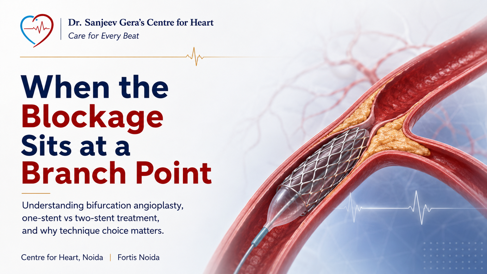
A coronary blockage is not difficult only because of how severe it is.
Sometimes, where the blockage sits matters even more.
One of the most challenging locations is a branch point, where one coronary artery divides into two vessels.
Patients may hear:
“The blockage is at a bifurcation.”
“A stent could affect the side branch.”
“This is more complicated than a routine angioplasty.”
“We may need one stent—or possibly two.”
This can understandably sound worrying.
However, a blockage at a branch point does not automatically mean that angioplasty cannot be performed. Modern interventional cardiology offers several strategies for treating these lesions.
The important part is choosing the right technique for that particular anatomy.
The coronary arteries supplying blood to the heart continually divide into smaller branches.
A bifurcation is simply a point where one main artery divides into two vessels.
Think of it like a road reaching a fork:
One road enters the junction → two roads leave it.
If a cholesterol plaque develops exactly at that junction, treating it becomes more complicated because the cardiologist must preserve blood flow through both important pathways.
This type of narrowing is called a coronary bifurcation lesion.
During a routine angioplasty, a stent is positioned across the narrowed segment and expanded.
At a bifurcation, placing a stent in one vessel can potentially affect the opening of the other vessel.
Imagine placing a scaffold inside the main road of a junction.
The scaffold may restore the main road perfectly—but part of it may lie across the entrance to the side road.
This creates several challenges.
The cardiologist needs to:
This is why bifurcation angioplasty requires more planning than a simple straight-vessel stent.
Modern coronary stents are made from a fine metallic mesh.
When a stent is placed across a bifurcation, some of its struts may naturally cross the entrance of the side branch.
In many cases, this is perfectly acceptable.
Blood can continue to flow through the spaces between the stent struts.
If necessary, the cardiologist can pass another guidewire through these openings into the side branch and further optimise the result.
The presence of stent struts across a side branch therefore does not automatically mean the branch has been blocked.
One important concern in bifurcation procedures is side-branch compromise.
When a balloon or stent expands inside the main vessel, several things can happen.
Part of the plaque can move toward the entrance of the side branch.
The natural dividing ridge between the two vessels—the carina—may move slightly toward one branch when the main vessel is expanded.
The inner lining of the artery can occasionally develop a tear around the side-branch opening.
The configuration of the stent at the branch opening can also influence blood flow.
Most of these situations can be managed during the procedure, but anticipating them is an important part of treatment planning.
A tiny branch supplying a small area of heart muscle is very different from a large branch supplying a major portion of the heart.
Before deciding how aggressively to protect a side branch, the cardiologist considers:
A larger, clinically important side branch deserves much greater attention.
A bifurcation can be thought of as having three important segments:
The artery before the branch point.
The continuation of the main artery after the split.
The second artery leaving the junction.
A blockage may involve one, two or all three of these segments.
That distribution strongly influences the treatment strategy.
Cardiologists sometimes describe a blockage as a true bifurcation lesion.
This generally means that significant disease affects both:
These lesions may require more planning because simply treating the main vessel may not adequately treat the side branch.
But even among true bifurcations, not every patient requires two stents.
This is one of the most common questions patients have.
The answer is:
It depends on the anatomy.
In many bifurcation lesions, cardiologists prefer to begin with a one-stent strategy, sometimes called a provisional stenting strategy.
Two stents are generally reserved for lesions where the side branch has significant disease or is unlikely to remain satisfactory after main-vessel treatment.
“Provisional” means that the cardiologist initially plans to place one stent in the main vessel while protecting the side branch with a guidewire.
If the side branch remains open with good blood flow, a second stent may not be necessary.
If the side branch develops significant narrowing or impaired flow, additional treatment can then be performed.
This approach avoids putting more metal into the artery than is necessary.
Although every procedure is individual, the broad process may look like this.
One guidewire is placed into the main vessel.
Another may be positioned in the side branch.
The side-branch wire acts as protection and makes it easier to access the branch if treatment becomes necessary.
Depending on the blockage, balloons or specialised plaque-modification equipment may be required before placing the stent.
If there is significant calcium, additional preparation becomes particularly important.
The stent usually extends from the vessel before the bifurcation into the main branch beyond the junction.
The stent may cross the opening of the side branch.
The cardiologist may use a balloon sized appropriately for the larger proximal part of the vessel.
This is an important step called the:
Coronary arteries often become smaller after they divide.
Therefore, the section of artery before a bifurcation is usually larger than the vessel beyond it.
A stent chosen to fit the smaller distal vessel may therefore not fully contact the vessel wall in the larger proximal portion.
During Proximal Optimisation Technique, a carefully sized short balloon is expanded in the proximal part of the stent.
This helps:
In modern bifurcation angioplasty, POT can be a very important technical step.
If the side branch maintains:
then additional intervention may not be necessary.
The procedure may end with just one stent.
This is often desirable because simpler stenting generally means:
But simplicity is useful only when the result is good.
If the side branch develops significant narrowing or reduced blood flow, the cardiologist may pass the wire back through the main stent into the side branch.
A balloon can then be used to open the branch.
If the result becomes satisfactory, a second stent may still be unnecessary.
If the branch remains significantly compromised, then conversion to a two-stent strategy may be appropriate.
In some bifurcation procedures, balloons are positioned simultaneously in:
The balloons are then inflated together at the junction.
Because they expand side by side, this is known as:
Kissing Balloon Inflation.
It can help reshape the bifurcation and improve the openings of both branches in selected situations.
It is not automatically required in every one-stent bifurcation procedure. Its use depends on the final anatomy and treatment strategy.
A planned two-stent technique may be considered when the side branch is:
In these situations, protecting only the main vessel may not provide an adequate result.
The cardiologist may therefore decide before the procedure that both vessels should be stented.
Because more stents do not automatically mean better treatment.
Two-stent procedures involve:
Therefore, two stents are generally used when the anatomy genuinely requires them.
The principle is:
Use the simplest technique capable of producing a safe and durable result.
There is no single two-stent method for every bifurcation.
Several techniques are available.
The choice depends heavily on:
Common techniques include the following.
A stent is placed in the side branch so that it meets the main-vessel stent approximately like the letter T.
This works best when the branch comes off at a favourable angle and precise placement is possible.
TAP is a modification of T-stenting.
The side-branch stent extends slightly into the main vessel to ensure that the branch opening is completely covered.
Additional balloon optimisation is then performed at the junction.
In the culotte technique, one stent is placed from the proximal main vessel into one branch.
A second stent then passes through the first stent into the other branch.
The proximal portion therefore contains overlapping stent layers.
The technique can provide good coverage when the two branches have relatively similar sizes.
With crush techniques, a portion of the side-branch stent is intentionally positioned within the main vessel and then compressed—or “crushed”—against the vessel wall by the main-vessel stent.
Several versions of this technique exist.
One of the best-known advanced strategies is:
DK Crush is a structured two-stent technique designed for certain complex bifurcation lesions.
It involves carefully positioning the side-branch stent, crushing its protruding section, rewiring the branch and performing balloon inflations at specific stages before and after the main-vessel stent is placed.
The “double kissing” part refers to balloon optimisation performed at two stages of the procedure.
DK Crush is particularly relevant in selected complex bifurcations, including some involving the left main coronary artery.
It is technically demanding and requires meticulous execution.
At a straight section of artery, small variations in stent position may have relatively limited consequences.
At a bifurcation, a few millimetres can matter enormously.
Incorrect positioning could potentially:
That is why bifurcation angioplasty is as much about planning geometry as simply expanding a blockage.
One important challenge is that the artery before the bifurcation is often larger than either branch after it.
For example, the proximal artery might be relatively large, while both daughter vessels are smaller.
If a stent is sized only to the larger proximal vessel, it may be too large for the distal branch.
If it is sized only to the smaller branch, it may initially be under-expanded proximally.
Modern bifurcation techniques account for this difference using carefully selected stent sizes and optimisation balloons.
The left main coronary artery is particularly important because it supplies blood to a very large proportion of the heart.
It usually divides into:
Some people also have an additional intermediate branch.
Disease at the distal left main bifurcation can therefore be especially significant.
Treatment may involve:
depending on the patient’s anatomy and overall condition.
When PCI is selected, detailed planning, appropriate bifurcation technique and intracoronary imaging become especially important.
No.
Some left main bifurcation lesions can be treated effectively with a one-stent provisional strategy.
Others have substantial disease extending into both the LAD and circumflex arteries and may be better suited to a planned two-stent approach.
The decision is based on anatomy rather than simply the label “left main disease.”
IVUS — Intravascular Ultrasound — allows the cardiologist to examine the coronary artery from inside.
Instead of relying only on a two-dimensional angiogram, IVUS can provide information about:
This can be especially valuable at complex branch points.
Optical Coherence Tomography (OCT) is another intracoronary imaging technology.
It provides extremely detailed images of the inside of the artery and stent.
Depending on the clinical situation, OCT may help assess:
The choice between different imaging techniques depends on the lesion and patient.
Some bifurcation lesions contain heavy calcium.
This creates additional challenges.
A rigid calcified artery may prevent a stent from expanding properly.
And at a bifurcation, incomplete expansion can compromise both the main vessel and side branch.
For selected heavily calcified lesions, the cardiologist may use plaque-modification technologies before placing the stent.
These can include techniques such as:
The appropriate method depends on the anatomy.
No.
An angiogram can sometimes make a side-branch narrowing appear more dramatic immediately after main-vessel stenting.
The important issue is whether the narrowing is actually restricting meaningful blood flow.
In selected situations, physiological measurements may help determine whether additional treatment is necessary.
This helps avoid putting in an unnecessary second stent purely because of how the angiogram looks.
The goal is not to create a perfect-looking photograph of both arteries.
The goal is to achieve:
Sometimes that requires one stent.
Sometimes it requires two.
The best technique is the one that appropriately matches the patient’s anatomy.
Before and during the procedure, the cardiologist considers:
A large branch is more important to preserve than a tiny branch.
A minimally diseased side branch may not require a stent.
A short narrowing at the opening is very different from disease extending far down the branch.
The angle affects how easily the artery can be accessed and which stent technique will work best.
Heavily calcified bifurcations may need additional lesion preparation.
The size and shape of the proximal and distal vessels influence stent selection.
A planned one-stent strategy can sometimes be converted to two stents if the side branch becomes compromised.
Bifurcation PCI can be more technically demanding.
Possible challenges include:
The actual risk varies greatly depending on the location and complexity of the lesion.
A simple bifurcation involving a small side branch is very different from a complex distal left-main bifurcation involving two large vessels.
The phrase does not necessarily mean that treatment is impossible.
It often means that treatment requires more planning than routine angioplasty.
Depending on the anatomy, options may include:
For highly complex disease, particularly involving multiple major arteries or the left main coronary artery, a multidisciplinary discussion may help determine whether angioplasty or bypass surgery offers the better overall treatment.
Not every complex bifurcation should automatically be treated with stents.
Bypass surgery may be preferable in some patients with:
The decision should therefore consider the entire coronary anatomy, not one branch point in isolation.
If you have been told that your blockage is at a branch point, useful questions include:
Which two arteries are involved?
How important is the side branch?
Is this considered a true bifurcation lesion?
Are you planning one stent or two?
Could the strategy change during the procedure?
How will the side branch be protected?
Will IVUS or OCT be used?
Is the artery heavily calcified?
Would bypass surgery provide a better result in my anatomy?
What is the expected benefit of treating this blockage?
Understanding the strategy can make the procedure much easier for patients and families to comprehend.
This is perhaps the most important message.
Patients sometimes assume:
One stent = simpler or incomplete treatment.
Two stents = more complete or advanced treatment.
That is not how bifurcation angioplasty should be judged.
If one well-positioned stent preserves excellent blood flow through both vessels, adding another stent may provide no additional advantage.
At the same time, when substantial disease involves an important side branch, deliberately using a well-planned two-stent technique may provide the better result.
The number of stents is not the goal.
The quality of the final result is.
A “70% blockage” or “90% blockage” tells only part of the story.
A cardiologist also needs to know:
Where is it?
A blockage in a straight segment of artery can be very different from the same percentage narrowing located exactly where two major vessels divide.
At a bifurcation, successful treatment depends on understanding:
That is why technique choice matters especially at branch points.
It is a coronary artery blockage located where one artery divides into two branches.
It can be more complex than routine angioplasty because the side branch must be protected. The level of risk depends greatly on the anatomy and the importance of the two branches.
No. Many bifurcation lesions can be treated with a one-stent provisional strategy.
Two stents may be considered when a large and important side branch has significant disease or is likely to remain compromised with a one-stent approach.
The side branch can often be rewired through the main-vessel stent and treated with a balloon or, when necessary, another stent.
It means treating the main vessel first with one stent while protecting the side branch and adding a second stent only if necessary.
DK Crush is an advanced planned two-stent technique used for selected complex coronary bifurcations.
It involves positioning balloons in both branches and inflating them together to optimise the shape and opening of the bifurcation in selected procedures.
IVUS provides detailed information about vessel size, plaque, calcium and stent expansion, helping optimise treatment in complex anatomy.
Selected left-main bifurcation lesions can be treated with PCI. However, bypass surgery may be more appropriate for some patients. The decision depends on the complete coronary anatomy and clinical situation.
Yes. A procedure may begin with a provisional one-stent strategy and convert to two stents if the side branch becomes significantly compromised.
Dr. Sanjeev Gera
MD, DNB (Cardiology), FSCAI
Centre for Heart, Noida
Fortis Noida
If you have been told that your coronary blockage is difficult because it lies at a branch point or bifurcation, the percentage of blockage alone does not determine the best treatment.
The size of both vessels, the length and distribution of disease, calcium, branch angle and overall coronary anatomy all influence whether treatment should involve medication, one-stent angioplasty, a planned two-stent technique or bypass surgery.
At a coronary bifurcation, the most important decision is not simply how many stents to use—it is choosing the right strategy for that specific artery.
 +(91)9810466173
 info@drsanjeevgeracardiologist.com