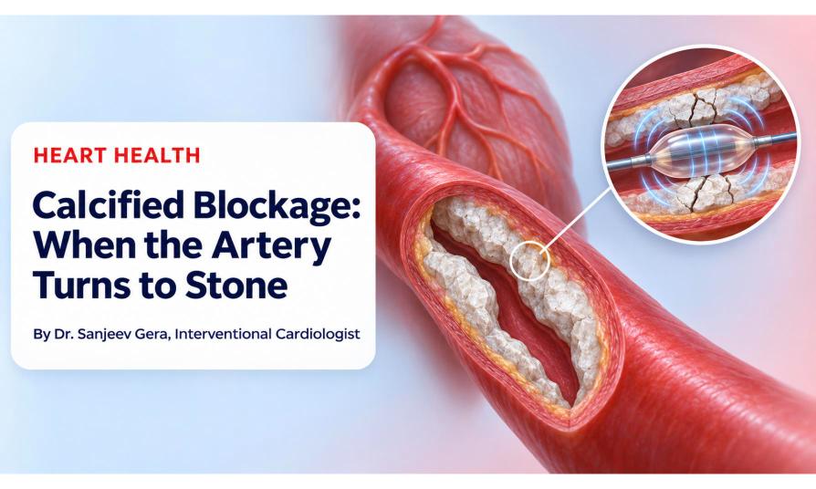
HEART HEALTH
By Dr. Sanjeev Gera, Interventional Cardiologist · 5 min read
Try pushing a soft balloon against a wall of stone. The balloon gives way long before the stone does.
That is the challenge of a calcified blockage, where calcium hardens the artery wall. Here is why a regular balloon can struggle, and how rotablation and shockwave (IVL) help.
Over the years, plaque in a heart artery can collect calcium and harden, much like bone. It becomes more common with age, diabetes and long-standing kidney disease.
Angioplasty relies on a balloon pushing plaque outward. Hard calcium resists that push, so the artery may not open well, and the stent placed afterwards may not expand fully.
A stent can only be as good
as the artery prepared for it.
Rotablation uses a tiny diamond-coated burr spinning at very high speed to shave hard calcium into microscopic particles, which pass safely through the circulation.
Intravascular lithotripsy (IVL), or shockwave, uses a balloon that releases sound-pressure waves. These crack the calcium in the artery wall, a little like breaking up kidney stones, so the artery can expand.
| Regular balloon | Rotablation | Shockwave (IVL) | |
|---|---|---|---|
| How it works | Pressure from inflation | High-speed burr shaves calcium | Sound waves crack calcium |
| Often suits | Softer plaque | Tight, heavily calcified narrowings | Deep calcium in the artery wall |
| Main limit | May not open hard calcium | Needs special training and care | The balloon must cross the narrowing |
Sometimes both are used in the same procedure. The right choice depends on your artery, not on one device being better for everyone.
Not always. Many calcified blockages can be treated with angioplasty after proper preparation. Your cardiologist will advise what suits you.
The area is numbed and you stay awake. Some people feel brief chest discomfort or, with IVL, a fluttering sensation. Tell the team about anything you feel.
No diet is known to dissolve it. A healthy lifestyle and medicines help protect your arteries from further damage.
No. Never stop them without asking your cardiologist.
Get your angiogram reviewed and understand your options.
This article is for general education and isn’t a substitute for personal medical advice. Please consult your cardiologist about your specific condition.
 +(91)9810466173
 info@drsanjeevgeracardiologist.com