HEART HEALTH
By Dr. Sanjeev Gera, Interventional Cardiologist · 5 min read
A map from above shows you where the road goes. It can’t show you the potholes. For that, you have to drive down the road itself.
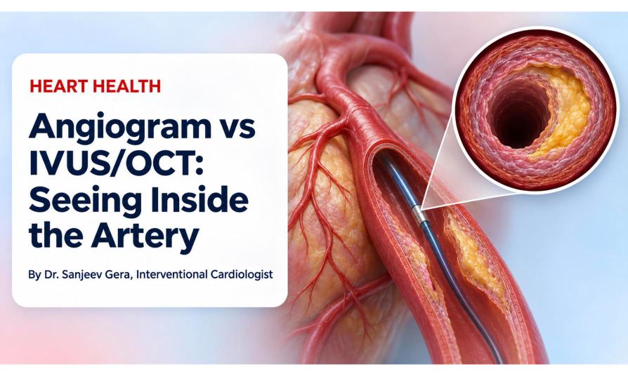
Heart imaging works the same way. An angiogram gives the outside view, while IVUS and OCT show the artery from the inside. Here is how they differ and why it matters.
Dye is injected into the heart arteries and X-rays record its flow. It is the standard test for finding blockages. However, it shows a shadow of the inside of the artery, not the wall where plaque actually builds up.
IVUS (intravascular ultrasound) places a tiny ultrasound probe inside the artery. It sees deep into the wall, which is useful for measuring vessel size and plaque.
OCT (optical coherence tomography) uses light to make extremely detailed pictures, which is useful for checking the stent and the artery’s inner surface.
The angiogram shows where to look.
IVUS and OCT show what is really there.
| Angiogram | IVUS | OCT | |
|---|---|---|---|
| View | Outline from outside | Cross-section from inside | Cross-section from inside |
| Uses | X-ray and dye | Sound waves | Light |
| Best for | Finding blockages | Depth, vessel size, plaque | Fine detail, stent check |
| When used | Almost every diagnosis | Selected or complex cases | Selected or complex cases |
No. They are especially useful in complex blockages, such as left main or calcified disease. Your cardiologist decides based on your case.
The test is done from inside the artery during your procedure and usually causes little or no extra discomfort.
They are established tools with a low risk in experienced hands. As with any procedure, your doctor will explain the risks.
Neither is better for everyone. Each has strengths, and the choice depends on your artery.
This article is for general education and isn’t a substitute for personal medical advice. Please consult your cardiologist about your specific condition.
 +(91)9810466173
 info@drsanjeevgeracardiologist.com