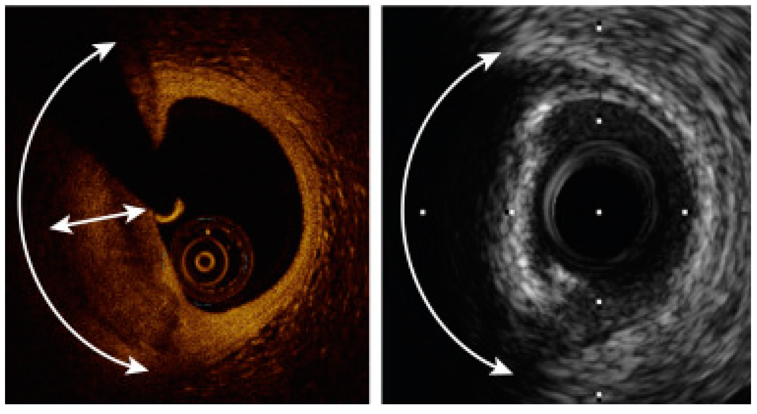
How IVUS and OCT Are Changing the Game in Heart Procedures
Imagine you’re fixing a leaky pipe in your home. You can guess where the leak is based on some damp spots on the floor, but wouldn’t it be easier if you had X-ray vision to see exactly what’s going on inside the pipe? That’s the kind of leap we’re seeing in the world of heart procedures, particularly with Percutaneous Transluminal Coronary Angioplasty (PTCA). Thanks to advanced imaging techniques like Intravascular Ultrasound (IVUS) and Optical Coherence Tomography (OCT), cardiologists now have a far clearer picture of what’s happening inside the coronary arteries during PTCA.
Let’s dive into how these technologies work and why they’re becoming a game-changer in treating coronary artery disease.
In the past, doctors performing PTCA had to rely primarily on angiograms—X-ray images of blood vessels—while navigating tiny catheters and balloons through the arteries. Though helpful, these images are 2D and don’t always tell the whole story. It’s like looking at a tree’s shadow instead of seeing the tree itself. But now, we have the tech to go beyond shadows.
Enter IVUS and OCT—two imaging tools that allow cardiologists to literally see inside the artery during a procedure. IVUS uses sound waves to create detailed cross-sectional images, while OCT uses light to provide high-definition visuals. Think of IVUS as an ultrasound for your heart’s arteries and OCT as a light-based scanner that can reveal intricate details.
Imagine you’re a cardiologist. You’re inside the coronary artery, about to place a stent—a tiny mesh tube that helps keep the artery open. But how do you know exactly where to put it and whether it’s going to stay in place? That’s where Intravascular Ultrasound (IVUS) comes in.
IVUS works by sending sound waves into the artery to generate 360-degree images of the blood vessel walls. These images allow doctors to:
IVUS is like a GPS for the heart, helping doctors navigate tricky procedures with greater precision.
While IVUS is amazing, sometimes we need even more detail. Enter Optical Coherence Tomography (OCT), which takes things up a notch by using light instead of sound. Imagine being able to zoom in and see individual leaves on a tree—OCT does something similar for the arteries. It’s so detailed that doctors can spot things as small as 10 microns (to put that into perspective, a human hair is about 70 microns thick).
With OCT, doctors can:
OCT’s high-resolution images provide a level of detail that was previously unimaginable, making it easier to customize treatments based on the patient’s specific needs.
You might be wondering, “So which is better, IVUS or OCT?” The truth is, it depends on the situation. They’re like different tools in a toolkit—each one excels in different scenarios.
In many cases, doctors may use both tools together to get the full picture—IVUS for the broad view and OCT for the fine details.
Let’s break it down. Why does all of this matter? Quite simply, it saves lives. Here are just a few of the benefits:
The future of imaging-guided PTCA looks bright. As technology advances, we can expect even clearer, more precise images that will continue to refine how doctors approach coronary interventions. The goal is simple: make procedures safer, more effective, and more personalized for each patient.
In the coming years, we’ll likely see even more integration of IVUS and OCT into standard care. And as more data comes in, the use of these imaging tools may become the gold standard for PTCA, helping doctors fix coronary arteries with surgical precision.
Conclusion
IVUS and OCT have revolutionized how cardiologists perform PTCA, turning it into a more precise, safer, and more effective procedure. With these advanced imaging tools, doctors can see what was once invisible, tailoring treatments to each patient’s unique anatomy and condition. It’s a new era for heart health—one where the odds are more in favor of the patient.
Stay tuned for more on the cutting-edge developments in coronary care, and remember, when it comes to your heart, precision makes all the difference.