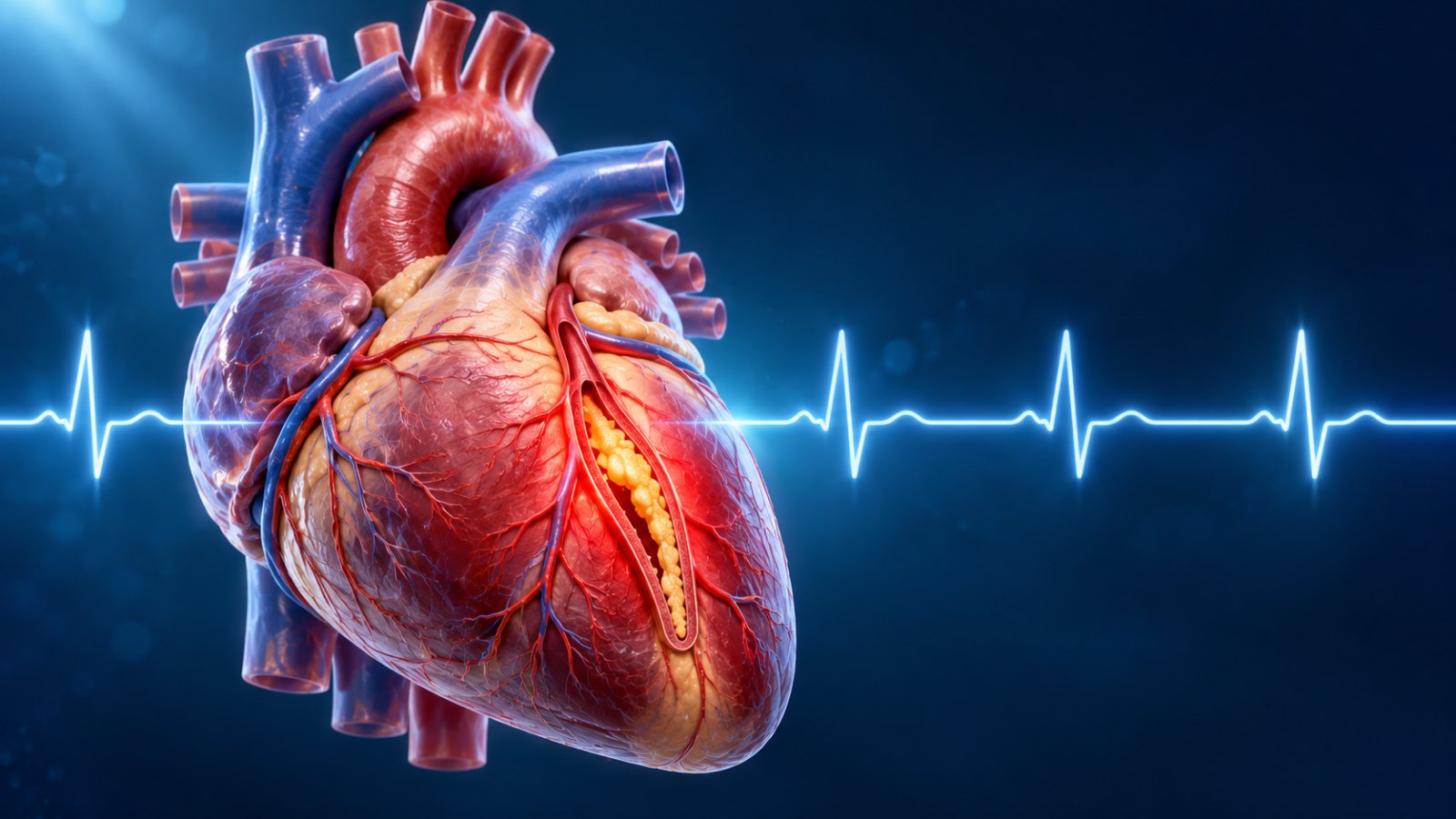
“Doctor, my ECG was normal just three months ago. How can I have a blockage?” It is one of the questions I hear most often in my Noida clinic, usually from someone sitting across from me with an angiography report they never expected to see.
The short answer surprises most people: a normal ECG does not mean your heart arteries are clear. An ECG is a brilliant, inexpensive, life-saving test, but it was never designed to “see” cholesterol building up inside your coronary arteries. Understanding what each heart test can and cannot tell you is one of the most useful things you can learn about protecting your heart.
Rakesh (name changed), a 46-year-old IT manager, had a “clean” ECG at his office health camp in spring. By August, he noticed a heaviness in his chest while climbing the metro stairs that faded the moment he stopped. He blamed acidity for weeks. A CT coronary angiogram finally showed an 85% narrowing in his main front artery. His ECG at rest, even on the day of diagnosis, looked completely normal.
An electrocardiogram (ECG or EKG) uses small stickers on your chest, arms and legs to record the electrical signals that make your heart beat. In about ten seconds, it tells a doctor a great deal: your heart rate and rhythm, whether there are conduction blocks, signs of an old heart attack, thickening of the heart muscle, and, crucially, whether the heart muscle is being starved of oxygen right now.
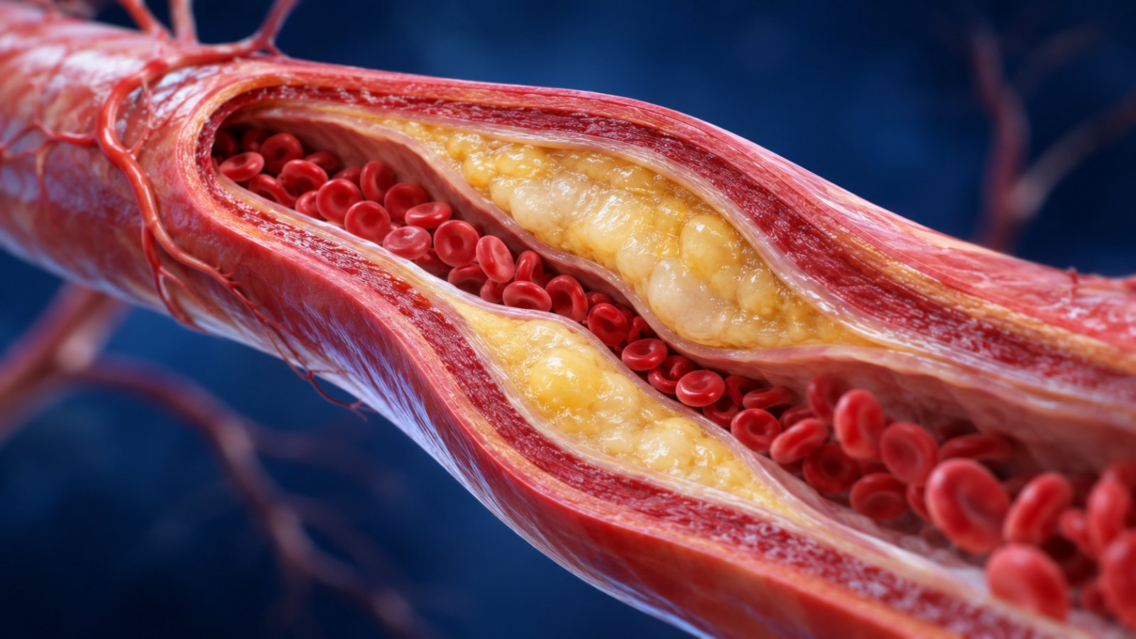
That last phrase is the key. Think of your coronary arteries as water pipes supplying a house. An ECG is like checking whether the taps are working. If the pipe is 70% clogged but enough water still trickles through while nobody is using it, the taps look fine. Only when everyone turns on the showers at once (when you climb stairs, walk briskly or feel stressed) does the shortage show up.
This is why a large share of patients with stable angina, the chest discomfort caused by narrowed arteries, have a normal resting ECG. The narrowing is real, but at rest the heart muscle still gets enough blood, so the electrical pattern stays normal.
An ECG tells you how your heart is behaving at this moment. It cannot tell you how much plaque is quietly building up in your arteries.
The single most important idea in this articleNarrowings of 50–90% often cause symptoms only when the heart works harder. Classic clues include chest tightness, heaviness, breathlessness, jaw or arm discomfort, or unusual fatigue that comes on with walking, climbing or emotional stress and settles within a few minutes of rest. If this pattern sounds familiar, a normal resting ECG should not reassure you.
During a heart attack, ECG changes can take time to appear, and some heart attacks never produce the dramatic “ST elevation” pattern at all. That is why emergency teams repeat the ECG every 15–30 minutes and check a blood test called high-sensitivity troponin, which detects heart muscle damage. If you have ongoing chest pain, sweating or breathlessness, a single normal ECG is never a reason to go home.
Many heart attacks occur when a plaque that was causing only a modest narrowing suddenly ruptures and a clot forms on top of it. These “soft” plaques cause no symptoms and no ECG change beforehand. They can, however, be seen on a CT coronary angiogram, which is why imaging has become so valuable in prevention.
No single test answers every question. Here is how cardiologists think about the most common ones, in plain language.
| Test | What it shows | What it can miss | Best used when |
|---|---|---|---|
| Resting ECG | Rhythm, heart rate, old heart attack, active oxygen shortage | Blockages that only matter on exertion; non-narrowing plaque | Every heart check-up and every episode of chest pain |
| 2D Echo | Heart pumping strength (EF), valves, wall movement, heart size | Mild-to-moderate blockages with normal pumping | Breathlessness, murmurs, suspected heart failure or valve disease |
| Treadmill test (TMT) | ECG changes and symptoms when the heart is pushed to work harder | Some significant blockages; false alarms are also common, especially in women | Exertional symptoms in people who can walk briskly |
| CT coronary angiography | A 3D picture of the arteries, including soft and calcified plaque | Precise severity in heavily calcified arteries | Chest pain with low-to-intermediate likelihood of disease; unclear stress tests |
| Coronary calcium score | Amount of hardened (calcified) plaque, as a risk number | Soft plaque without calcium; not a test for current symptoms | Risk assessment in people without symptoms, e.g. deciding on statins |
| Coronary angiography (cath) | The gold-standard view of blockages, with option to treat in the same sitting | Little, but it is invasive | High suspicion, abnormal non-invasive tests, or a heart attack |
Modern international guidelines recommend CT coronary angiography as an early test for many people with suspected angina. It takes a few minutes, needs only an IV injection of contrast, and a normal scan is highly reliable at ruling out significant blockage. It also detects early plaque that would never show up on an ECG or a treadmill test, giving you a chance to act years before a heart attack.
Yes, in the right person. It shows how your heart responds to effort, how fit you are, and whether symptoms appear at a particular workload. But it is not perfect: it can miss some significant blockages and produce false-positive results, so it should be interpreted alongside your symptoms and risk factors rather than in isolation.
You deserve a more detailed heart evaluation, even with a normal ECG, if any of the following apply:
Indians tend to develop coronary artery disease roughly a decade earlier than people in Western countries, often with more extensive disease. In my practice, heart attacks in people in their 30s and early 40s are no longer rare. Relying on a yearly ECG alone is simply not enough for this group.
Call emergency services or go to the nearest hospital with a cath lab immediately. In a heart attack, every minute counts. Learn more about the warning signs your heart may be giving you.
Because his narrowing was found before it caused a heart attack, Rakesh had a planned angioplasty with a single stent, guided by intravascular imaging. His heart muscle was undamaged. He went home the next day and was back to his morning walks within a week, now with his cholesterol, blood pressure and blood sugar under tight control. (Not every narrowing needs a stent, though. Read 70% blocked artery: do you actually need a stent?)
The lesson from his story is not that ECGs are useless. They are essential. The lesson is that your symptoms and your risk factors deserve as much attention as your reports. When something feels different, trust that feeling and get it properly evaluated.
Not directly. An ECG can show signs that the heart muscle is short of oxygen, or that an old heart attack has occurred, but many significant blockages leave the resting ECG completely normal. Tests such as CT coronary angiography, a stress test or a coronary angiogram are needed to look for blockages.
Yes. In the early stages of a heart attack, and in some types of heart attack, the ECG may be normal or show only subtle changes. That is why hospitals repeat the ECG and measure troponin levels before ruling out a heart attack.
They answer different questions. A TMT shows how your heart responds to exercise, while CT coronary angiography shows the arteries themselves, including early plaque. For many people with suspected angina, CT angiography is now preferred as the first test, but the right choice depends on your age, symptoms, kidney function and heart rate. Your cardiologist will advise you.
For healthy adults, an ECG is usually part of a routine check-up. If you have diabetes, high blood pressure, high cholesterol or a strong family history, your cardiologist may recommend periodic ECGs alongside other tests tailored to your risk.
No. An echo is excellent for assessing heart pumping and valves, but people with significant blockages often have a normal echo at rest, especially if they have never had a heart attack.
It is a quick, widely used test with low radiation on modern scanners. It uses an iodine-based contrast dye, so your kidney function and any allergies are checked beforehand. It is not suitable for everyone, for example those with very irregular or very fast heart rates, so it should be ordered by a doctor.
If your reports say “normal” but your body is telling you something else, let’s find out together. Dr. Sanjeev Gera offers evidence-based heart evaluations in Noida, with treatment at Fortis Hospital, Noida.
Fortis Hospital, Sector 62, Noida (Mon–Sat, 8:30 AM–5:30 PM) · Center for Heart, Spectrum Mall, Sector 75 (Mon–Sat, 7:00–9:30 PM)This article is for general education and does not replace a personal medical consultation. If you have chest pain or other urgent symptoms, seek emergency care immediately.
 +(91)9810466173
 info@drsanjeevgeracardiologist.com