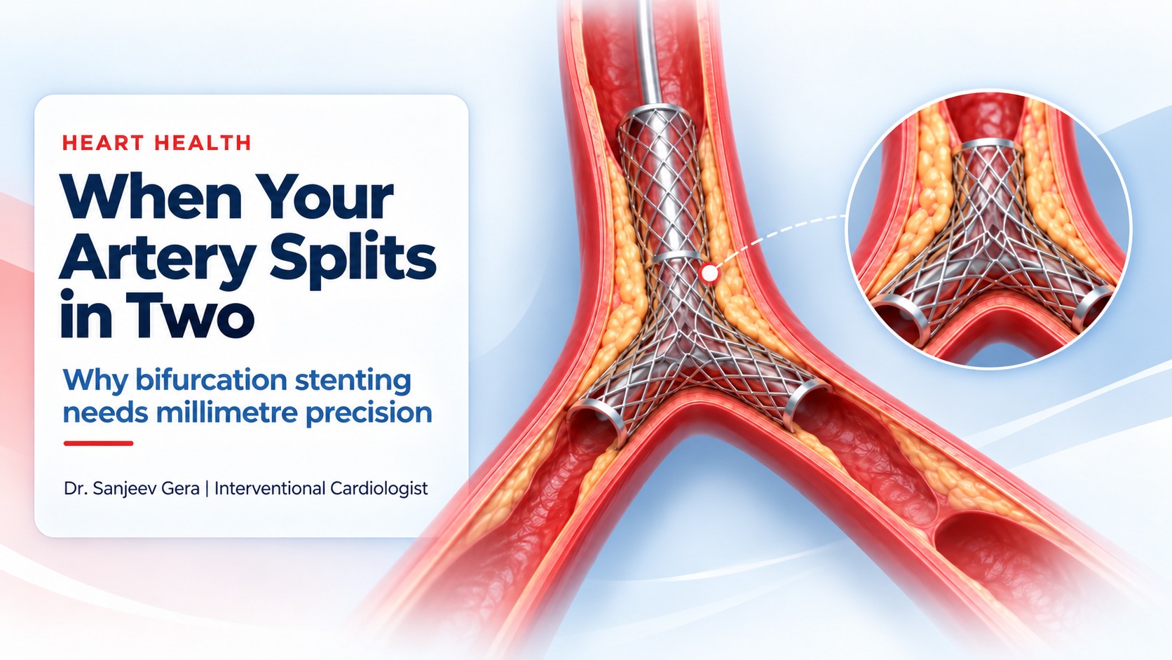
Picture a highway that suddenly forks into two roads, with a traffic jam sitting right at the fork. Clear one lane carelessly and you could block the other.
That is exactly the challenge cardiologists face with a bifurcation blockage in the heart. Here’s what it is, how it differs from a regular stent, and why millimetre precision matters.
A stent is a tiny mesh tube, usually made of a cobalt-chromium or platinum-chromium alloy and coated with a drug that stops scar tissue from regrowing.
It travels to the narrowed artery on a thin balloon, usually through the wrist. When the balloon inflates, the stent opens, presses the plaque against the artery wall, and stays there as a permanent scaffold so blood can flow freely again.
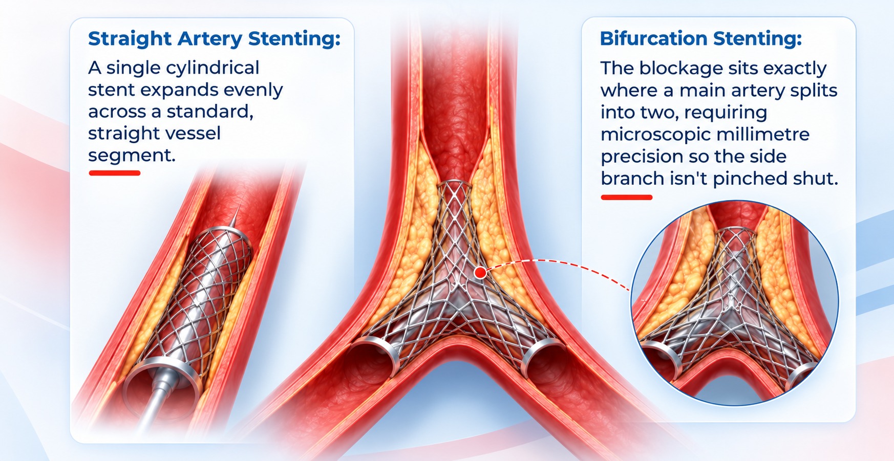
The blockage sits in a straight segment. One cylindrical stent expands evenly across it. Quick and predictable: one fix, one lane, done.
The blockage sits exactly where the artery splits. The stent must be placed with millimetre precision so the side branch isn’t pinched shut.
Your coronary arteries branch like a tree. Turbulent blood flow at these forks makes them a favourite spot for plaque to build up.
When a stent opens in the main artery, plaque can be pushed sideways (called plaque shift), and the metal struts can lie across the mouth of the side branch. Done without care, this can pinch the side branch shut, and that branch may supply an important part of your heart muscle.
The goal isn’t just to open one artery.
It’s to keep both branches of the fork healthy.
Angiography, often with IVUS or OCT imaging from inside the artery, measures each branch, the plaque and the angle between them.
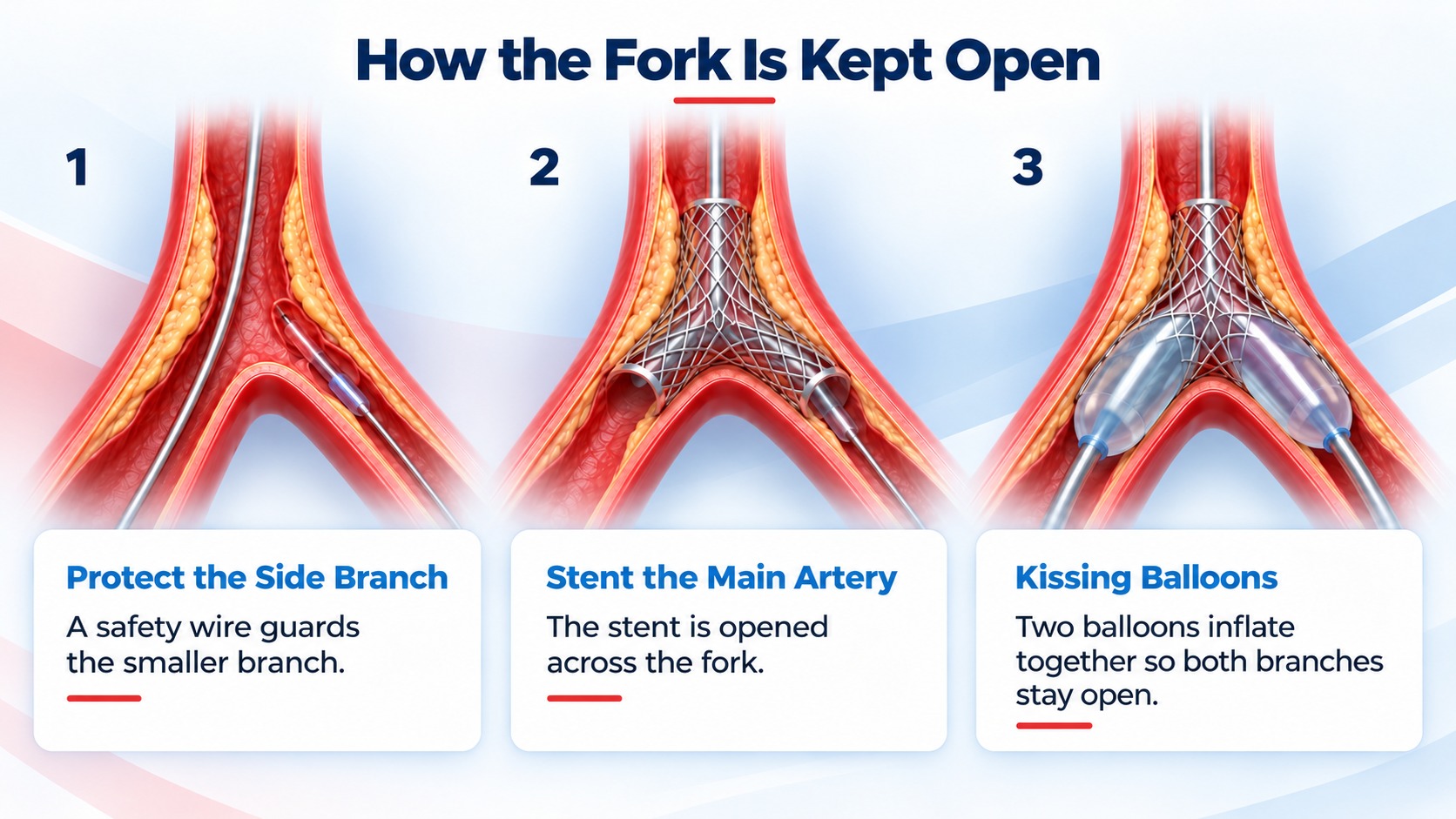
One stent with a safety wire in the side branch suits most cases. Two stents (DK-crush, culotte, T-stenting) are used when both branches are diseased.
The proximal optimisation technique (POT) and kissing balloon inflation shape the stent so both branches stay wide open.
| Straight artery | Bifurcation | |
|---|---|---|
| Location | Straight vessel segment | Where an artery splits in two |
| Stents needed | One | One or two, depending on anatomy |
| Main risk | Re-narrowing | Pinching the side branch |
| Extra techniques | Rarely needed | Side-branch wire, POT, kissing balloon, imaging |
| Complexity | Standard | Higher, needs careful planning |
An angiogram confirms where the blockage is, and your doctor explains the planned approach.
Usually done under local anaesthesia through the wrist. Fork cases may take a little longer.
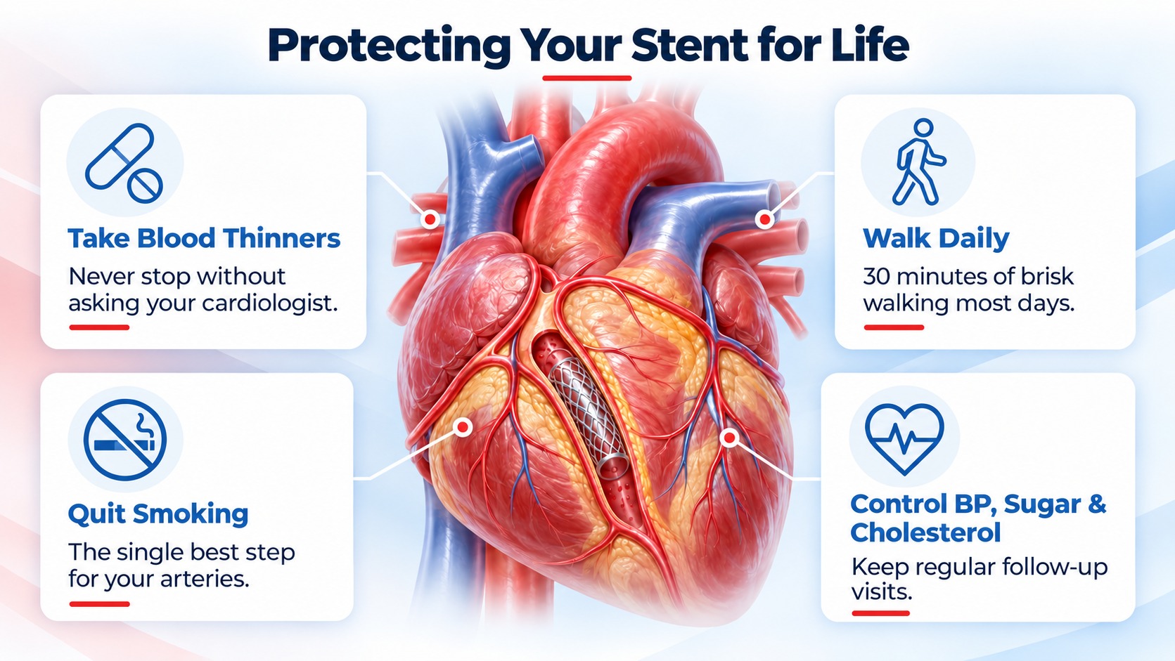
Most patients go home in a day or two, on blood thinners taken exactly as prescribed.
It is technically more complex, so planning and experience matter. With modern imaging and techniques, results in experienced hands are excellent.
No. Most bifurcations are treated with a single stent while the side branch is protected. A second stent is used only when needed.
No. Stopping them early can cause the stent to clot. Always speak to your cardiologist before changing any medicine.
Many patients resume light activity within days. Your cardiologist will guide you on exercise, driving and work.
Chest pain, breathlessness on exertion, or a family history of heart disease? Get your heart checked and understand your options.
This article is for general education and isn’t a substitute for personal medical advice. Please consult your cardiologist about your specific condition.
 +(91)9810466173
 info@drsanjeevgeracardiologist.com